
Anterior Cervical Discectomy With Fusion
Anterior cervical fusion is an operation performed on the upper spine to relieve pressure on one or more nerve roots, or on the spinal cord. The term is derived from the words anterior (front), cervical (neck), and fusion (joining the vertebrae with a bone graft).
Click Here to Download Brochure
Why Is It Done?
When an intervertebral disc ruptures in the cervical spine, it puts pressure on one or more nerve roots (often called nerve root compression) or on the spinal cord, causing pain and other symptoms in the neck, arms, and even legs. In this operation, the surgeon reaches the cervical spine through a small incision in the front of the neck. After the muscles of the spine are spread, the intervertebral disc is removed and a bone graft is placed between the two vertebral bodies. Over time, this bone graft will create a fusion between the vertebrae it lies between.
In more than ninety percent of cervical spine fusion surgeries done today a small cervical plate is used to stabilise the spine immediately after surgery. This hardware is used to improve the stability of the spine immediately after surgery and to also decrease the chance that the bone graft might be dislodged or moved slightly from the position that it was placed in by the surgeon. The use of hardware for stabilising the cervical spine has changed the way in which cervical collars are used after surgery. Today, collars are typically worn for a shorter period of time after surgery than in the past.
What Happens Afterwards?
Successful recovery from anterior cervical fusion requires that you approach the operation and recovery period with confidence based on a thorough understanding of the process. Mr. D'Urso has the training and expertise to correct physical defects by performing the operation; he and the rest of the health care team will support your recovery. Your body is able to heal the involved muscle, nerve, and bone tissues. Full recovery, however, will also depend on you having a strong, positive attitude, setting small goals for improvement, and working steadily to accomplish each goal.
The Operation
Incision
Surgery for anterior cervical fusion is performed with the patient lying on his or her back. A small incision is made in the front of the neck, to one side.
Exposure and Removal of the Cervical Disc
After a retractor is used to pull aside fat and muscle, the disc is exposed between the vertebrae. Part of it is removed with a forceps.
Then a surgical drill is used to enlarge the disc space, making it easier for the surgeon to empty the intervertebral space fully and remove any bone spurs. Afterwards, only a single ligament separates the surgical instruments from the spinal cord and nerve roots.
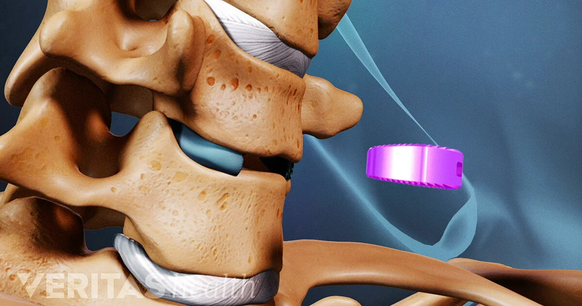
Placement of the Fusion Cage
A biocompatible plastic (PEEK) cage filled with tricalcium phosphate bone substitute in then placed into the space. This is a new alternative Mr. D'Urso uses instead of a traditional bone graft harvested from the iliac crest (pelvis). This avoids a second incision and aids a faster recovery. New bone grows through the cage to allow a rapid fusion to the adjacent vertebrae to occur.
Adding Stability: Fusion
Placing a fusion cage between the two vertebral bodies is done in order to create a fusion between these bones. The fusion is a direct result of the fusion cage, but small, specialised metal plates are also placed on the front of the cervical spine in order to increase the stability of the spine immediately after the operation. Surgeons use cervical hardware to decrease the amount of time that you will have to wear a collar after surgery, and also to increase your chances of getting a solid fusion between the two vertebral bodies. As the cage is filled with a bone substitute material, this also assists the fusion process.
Incision Closure
The operation is completed when the neck incision is closed in several layers. Unless dissolving suture material is used, the skin sutures (stitches) or staples will have to be removed after the incision has healed.